| 会员中心 | 设为首页 | 添加收藏 |
 |
| 当前位置:首页 > 供应信息 > 产品信息 > |
小动物PETCT:小动物PET/MRI成像系统, |
更新时间:2026-11-16 04:07:31 |
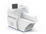
小动物PETCT小动物PET/CT的用途 小动物PET/CT是当今全球医药领域及生物工程方面的领先技术,通过对小型啮齿动物(小鼠或大鼠)进行活体状况下的功能及解剖成像,获得该动物身体状况及药物在小动物体内分布情况的各种数据。能对肿瘤、神经、心血管等疾病以及遗传基因研究,药物临床前筛选等提供先进的技术支持。 随着基因组学的发展,小动物在现代分子生物学实验中日趋重要。高分辨率小动物PET出现,成为分子显像的重要工具。基因表达、基因治疗及药物研究需要活体内分子显像。小动物PET可以进行肿瘤代谢显像、受体显像、基因表达显像等,是一种活体的功能显像。CT不仅可以给PET提供精确的衰减校正和解剖定位,而且可以提供形态学的诊断信息,可以得到更全面更准确的信息。能够提供丰富解剖细节的CT与具有功能显像的PET相结合是分子影像发展的潮流方向。 小动物PET/CT的特性 与其他分子显像方法相比具有以下优点: 1.可动态地获得秒数量级的动力学资料,能够对生理和药理过程进行快速显像。 2.具有很高的空间分辨率,可以观测到小鼠心血管、肿瘤组织形态等细微结构。 3.具有很高的灵敏度。 4.是一种相对无创伤性方法,示踪量的PET显像剂不会产生明显的药理毒副作用,活体CT具有低 剂量的特点、不会产生辐射伤害。 5.小动物PET扫描仪体积小、结构紧凑、空间分辨率高。6.使用活体小动物PET/CT影像可大幅度减少实验费用。 小动物PET/CT的放射性核素 小动物PET/CT是根据临床正电子发射/计算机辅助断层扫描成像技术原理设计,成像时所需的射线来自注射入动物体内的放射性核素。可用的放射性核素有18F、11C、13N、15O、68Ga、82Rb等,常用的主要是18F和11C 。 小动物PET/CT的应用领域 小动物PET/CT在肿瘤、神经科学研究、心脏病学、临床前药物研究及遗传 基因研究领域中应用广泛。 1、在肿瘤研究中的应用 2、在神经科学研究中的应用 3、在心脏病学研究中的应用 4、在临床前药物研究中的应用 5、在遗传基因研究中的应用小动物PET/MRI成像系统,深圳中小学创新实验室 宝诺集团 吊装式物理实验室-MOTELAB宝诺集团进口频谱分析仪USB端口 吊装式化学实验室-MOTELAB宝诺集团吊装式生物实验室 宝诺集团 |
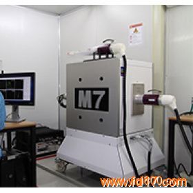
小动物PET/MRI成像系统,M系列小动物磁共振成像系统是Aspectimaging公司推出的专用的小动物MRI,采用高性能钕铁硼永磁体,实现高分辨率的小动物核磁共振成像。 M系列分为,M3,M7,M12,MRI-PET几个型号,除用于常见的大鼠和小鼠成像外,也可定制用于兔子,猴,小型狗等实验动物的核磁共振活体成像。还可以原机增加PET线圈,升级成PET/MRI,PET分辨率达到0.8mm. 相比于传统的超导式小动物MRI,永磁式M系列小动物MRI有明显的现实优点: 1、价格只有1/3甚至更低 2 、免去新建核磁屏障室的麻烦,普通实验室即可 3 、M系列运转消耗为零,比传统磁共振每年可结省几十万的运转消耗费 4 、更好的磁共振造影剂T1成像效果 M系列小动物磁共振是一款小巧紧凑桌上型microMRI,容易摆放。M系列小动物核磁共振系统平台完全满足最严格的安全要求标准,可以确保实现无边缘磁场、零外场强度,因此无需任何特殊外围防护/屏蔽室或其他操作规程限制。 M7 Compact MRI for DedicatedMouse and Rat Imaging The M7 compact, high-performance, one-touchMRI system is intended for dedicated imaging of mice and rats weighing up to700 grams. The M7 enables 2D and 3D, ex vivo, in vivo and in vitro imaging. Thesystem can be uniquely placed behind the bio-containment barrier for in vivoimaging. In addition to its comprehensive suite of applications for mousephenotyping, Aspect Imaging’s LumiQuant? package for generation of 3Dtomographic quantification of luminescence and co-registration with compact MRis optimized for use on the M7 platform. Click here to read more aboutM7 at the MIC, USC. One instrument, multipleapplications The M7 has all the capabilities to routinelyand repeatedly quantify phenotypes in rats and mouse models of disease,including: ·Cancer Research ·Cardiovascular ·Contrast-based Molecular Imaging ·Diabetes and Obesity ·Embryology / Developmental Biology ·Nephrology ·Neurobiology ·Stem Cell / Cell Tracking 3D Whole Body MorphologicalImaging The M7 compact, high-performance MRI systemenables high-resolution 3D whole body morphological imaging of rat and mousemodels of disease. Biologists and researchers without prior MR or imagingexperience can now leverage the gold standard modality – MRI – for morphological imaging. The M7is an in vivo imaging platform that is: ·Easy to learn, ·Effortless to operate, and ·Fast to generate reproducible, quantitativeresults of phenotypes and therapeutic ·efficacy. Maintenance-free The M7 provides a high-performance in vivoMRI system that is virtually maintenance free. The system requires: ·No cryogens, ·No auxiliary power, and ·No compressors or plumbing that requireservicing Infrastructure-free The M7, like all compact MRI systems fromAspect Imaging, can be placed in most locations in a facility and doesn’trequire an infrastructure typically needed with superconducting MRI system orcryogen-free MRI systems. The system therefore does not require any of thefollowing: ·No shielding ·No auxiliary power supplies or services ·No plumbing, pumps or compressors The M7 can therefore be placed: ·In a small animal core imaging facility ·Right next to other modalities like PET, CTor optical ·In a working lab ·Behind the barrier 应用: 应用于肿瘤生长,肿瘤转移成像,神经生物学,脑血管病,心血管学,生殖与发育,糖尿病与肥胖,解剖和组织胚胎学,磁共振造影剂等分子影像领域的小动物活体成像研究,并可以通过DICOM与microPET,小动物SPECT,micro-CT,小动物超声,小动物活体荧光,生物发光等多种小动物活体成像技术进行多模态融合成像。 |
 |
热门产品 |  |
 |







|